Lung lesion analysis is, through some distance, the ideas maximum steadily amassed, principally to substantiate and quantify breathing issues, in addition to to evaluate the result of positive intervention methods. The 2 maximum prevalent pulmonary lesions noticed on the pig slaughterhouse are:
- Craneo-ventral pulmonary consolidation (CVPC)
- Pleuritis, basically in caudal lobes.
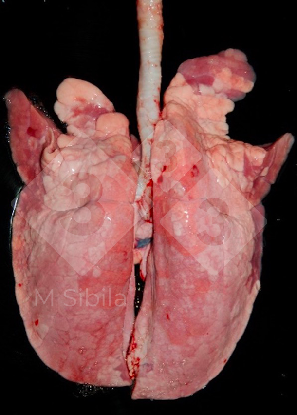
Within the provide article, CVPC refers to purple-dark and well-defined spaces of pulmonary consolidation, basically positioned bilaterally in apical, intermediate, accent in addition to the cranial a part of the diaphragmatic lung lobes (Determine 1).


Generally, this sort of lesion is related to Mycoplasma hyopneumoniae (M. hyopneumoniae) an infection. Alternatively, this lesion may also be produced through different pathogens, comparable to swine influenza virus in a multifocal trend, and Bordetella bronchiseptica and Pasteurella multocida in a extra diffuse method. Due to this fact, the causative agent(s) will have to be decided by the use of laboratory ways.
In acute M. hyopneumoniae illness instances (and with out secondary infections), the lesion is apparent and well-demarcated, being simply identified and scored. Alternatively, normally, the lesion is annoyed with the presence of different bacterial or viral pathogens, various in colour, consistency, and extension (however now not the craneo-ventral location).
At slaughterhouse, CVPC lesions have a tendency to be power, the colour has a tendency to be greyish, and the tissue may also be retracted forming scars or interlobular fissures, complicating its remark and scoring. If different lesions are provide, comparable to pleuritis, the scoring from the affected lobes may even be tougher or now not imaginable to evaluate. Macroscopically the severity of CVPC is measured through its extension; the upper the share of affected tissue, the more serious the lesion.
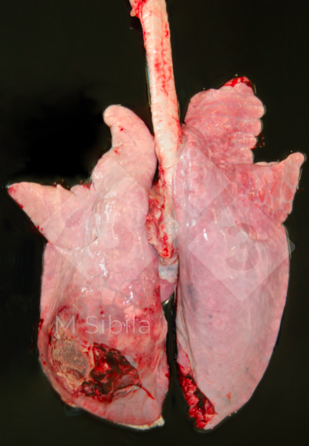
Pleuritis refers back to the irritation of the pleural serosa. When this lesion is confined to dorso-caudal lobes, it’s strongly suggestive of Actinobacillus pleuropneumoniae an infection (Determine 2). Continual instances (the ones normally provide at slaughterhouse) are characterised through fibrous adhesions to visceral and parietal pleura. In such instances, lung tissue adhesions are not unusual and depart a part of the organ adhered to the thoracic wall right through the lung’s elimination from carcass, resulting in its condemnation. On this state of affairs, presence of those lung adherences can be indicative of top severity.

Lung lesion scoring methods are simple and non-invasive strategies that supply knowledge on occurrence and extension (however now not occurrence) in a moderately reasonably priced (no further subject material is wanted) means. Alternatively, it’s also a non-confirmatory (no etiologic prognosis) and subjective (coaching is wanted) estimation, that can be tricky to arrange (particularly when pigs are despatched to slaughterhouse in different vans or when surprising adjustments at the arrival or slaughtering time occur) and, as a result, turns into pricey and time-consuming for evaluators.
There’s a plethora of CVPC lung lesion scoring methods, maximum of them in keeping with a visible estimation of the lung tissue affected (in issues or percentages) (Desk 1). Different methods use a three-d means through normalizing the share of the lung tissue suffering from the relative weight or quantity of every lobe. Irrespective of the variations, a excellent correlation a few of the primary CVPC scoring strategies maximum steadily used at slaughterhouse used to be demonstrated. Some scoring methods use diagrams or footage to assist to report the lesions, permitting a retrospective and actual research however making them unpractical because the plucks go back and forth extraordinarily speedy throughout the slaughter line.
Desk 1. Primary craneo-ventral pulmonary consolidation (CVPC) scoring methods (tailored from Maes et al., 2023).
| Manner | Gadgets | General ranking | Parameter used to attain the lesion at lobe degree |
|---|---|---|---|
| Goodwin et al., 1969 | issues | 0-55 | lesion trend |
| Hannan et al., 1982 | issues | 0-35 | measurement of the lobe |
| Madec and Kobisch (1982) | issues | 0-28 | 4 issues in line with lobe |
| Morrison et al., 1985 | proportion | 0-100 | weight of the lobe |
| Straw et al., 1986 | proportion | 0-100 | measurement of the lobe |
| Christensen et al., 1999 | proportion | 0-100 | weight of the lobe and lesion trend |
| Ph. Eur. approach | proportion | 0-100 | weight of the lobe |
| Sibila et al., 2014 | proportion | 0-100 | delimitation in a picture of the realm of affected tissue |
In a similar way, there are a number of scoring methods for pleuritis (Desk 2).
Desk 2. Pleuritis scoring methods for use at slaughterhouse (tailored from Maes et al., 2023).
| Manner | Primary parameter | Ranking | Classification |
|---|---|---|---|
| Madec and Kobisch (1982) | Localization, extension and measurement (diameter) of the pleuritic lesion | Ranking 0-4 in line with lobe. General 28 issues. | 0: No pleuritis 1: Interlobular pleuritis 2: Localized pleuritis <2cm 3: Intensive pleuritis <2cm diameter with adhesions to ribcage 4: Partial or general ribcage condemnation |
| CTPA Pagot et al., 2007 |
Extension and chronicity of the pleuritic lesion | 0-2 | 0: No pleuritis 1: Fibrinous pleuritis 2: Prolonged pleuritis: lungs can’t be got rid of from the carcass |
| Pointon et al., 1992 |
Form of adhesions and presence of pneumonia | 0-2 |
0: No pleuritis
2: Pleuritis (adhesions of lungs to chest wall):
|
| SPES Dottori et al., 2007 |
Localization, extension of the pleuritic lesion | Ranking 1-4 in line with lobe. General 28 issues. | 1: Craneo-ventral lesion: interlobar pleuritis or at ventral border of caudal lobes 2: Dorso-caudal monoliteral focal lesion 3: Bilateral lesion of sort 2 or prolonged monoliteral lesions (no less than one 3rd of 1 diaphragmatic lobe) 4: A number of prolonged bilateral lesion (no less than one 3rd of 1 diaphragmatic lobe) |
CTPA: Gadget through the Centre Method de Productions Animales
SPES: Slaughterhouse Pleurisy Analysis Gadget
- The gadget described through Madec and Kobisch (1982) ranking pleuritis lesion from 0 to 4, giving the easiest ranking when partial or general ribcage is condemned because of lung adherences.
- Slaughterhouse Pleurisy Analysis Gadget (SPES) additionally ratings the pleuritis lesions from 0 to 4 however making an allowance for their localization, extension and measurement (diameter).
- The Centre Method des Productions Animales (CTPA) approach handiest differentiates between fibrinous pleuritis amongst lung lobes from the ones pleuritis inflicting lung adherences to the ribcage.
- The Pointon gadget (Pointon et al.,1992) separates the interlobular pleuritis from those adhered to the ribcage additionally making an allowance for the presence of pneumonia.
The choice of the scoring gadget to be carried out on the slaughterhouse will have to be decided relying on:
- slaughter line traits: velocity, flowchart and accessibility
- permissibility of the slaughterhouse to gather samples (for extra actual scoring or for diagnostic affirmation)
- form of lesions to be scored
- selection of animals to be assessed
- team of workers sources to be had to accomplish such scoring.
The usage of voice recording to check in the lesion ranking could also be of significant assist to counteract the highspeed line permitting the handbook palpation of the lungs. Synthetic intelligence-based the best way to robotically overview lung lesions would possibly assist to automatize and objectivize the method. Alternatively, those methods are nonetheless beneath building as they want to be skilled and tailored to seize and analyze the picture from a dangling and shifting pluck of lungs.
Transient abstract of the thing: Evaluation at the technique to evaluate breathing tract lesions in pigs and their manufacturing affect. Maes D, Sibila M, Pieters M, Haesebrouck F, Segalés J, de Oliveira LG. Vet Res. 2023 54(1):8. doi: 10.1186/s13567-023-01136-2.
