
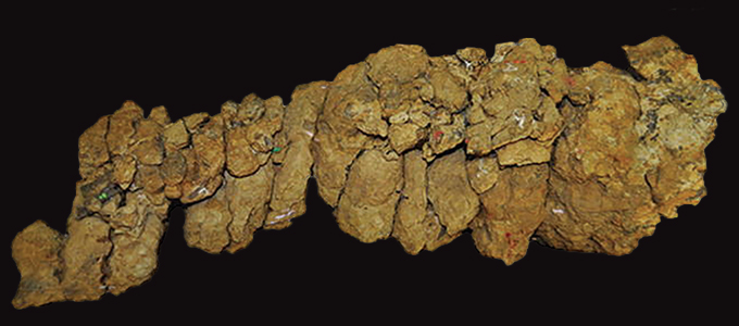
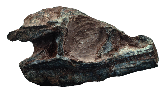
Shattered crocodile. Officially, Confractosuchus. It was once found out in Australia when a bulldozer clearing a boulder broke a stone into items. Uncovered parts of the broken-up rock made transparent that fossils had been inside of, however there was once no quick signal that this discovery would later disclose an exceptional snapshot of lifestyles from the Cretaceous Length.
Paleontologist Matt White of the College of New England in Armidale, Australia, and co-workers organized to have the fossil-laden rock scanned with X-ray computed tomography. Like a clinical CT scan, the process takes a couple of photographs of an object that may be assembled right into a three-D map of the inner. The workforce was hoping to make use of the scans as guides to isolate particular person bones within the fossil with out taking out them, then manipulate the three-D photographs to nearly put the shattered croc again in combination.
However one phase of the fossil puzzle gave them bother. Iron-rich stone surrounding the bones made it tricky to get excellent X-ray photographs. So the researchers determined to take a look at some other manner.
They despatched the thriller bite to chemist Joseph Bevitt of the Australian Centre for Neutron Scattering in Sydney, who focuses on the use of subatomic neutron debris to symbol historic gadgets. Together with the predicted croc bones, Bevitt found out one who appeared like a dinosaur leg bone. It was once within the portion of rock the place the crocodile’s abdomen hollow space would had been.
“Once I noticed the neutron consequence and the little dino femur, I used to be shaking with surprise,” Bevitt says, “each in awe and doubt with what we had viewed.”
Years of study plus extra X-ray and neutron scanning in the end showed that the stays of a in the past unknown species of dinosaur, bitten into chunks and scored with teeth marks, had been within the croc’s abdominal. The discovering earned the shattered crocodile the second one part of its title: sauroktonos, for lizard killer. White, Bevitt and co-workers revealed their discovery of each the newly known species of crocodile and the never-before-seen dinosaur inside of closing yr in Gondwana Analysis (SN: 3/26/22, p. 5).
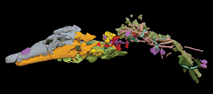
It’s a surprising discovery: Confractosuchus sauroktonos, the shattered crocodile lizard killer, and the stays of its closing meal, its dinosaur sufferer, frozen in stone 100 million years in the past. It’s a vignette that can by no means have come to gentle if no longer for neutron tomography. Even supposing neutrons had been used for imaging in business and armed forces packages since in a while after the neutron was once found out in 1932, it’s handiest in the previous couple of a long time that those subatomic debris have begun to supply scientists with exceptional perspectives inside of fossils and antiquities.
Glance, don’t contact
There was once a time when learning fossils and artifacts steadily intended destructive or destroying them. Mummified stays had been dissected. Sealed boxes had been cracked open. Fossils had been pried free from rock. In some instances, fossil-containing samples had been floor down, layer by means of layer, to create photographs of sequential parts in slices that exposed the fossilized constructions inside of.
Thankfully, X-rays be offering nondestructive insights. As a high-energy type of electromagnetic radiation, or gentle, X-rays have interaction with the electrical and magnetic fields related to electrically charged debris. In a physician’s workplace, when a technician shines a beam of X-rays at a damaged leg, the sunshine will get scattered or absorbed by means of the fields of the electrons round atoms within the leg. The denser a subject material is, the extra electrons are packed in it, and the fewer successfully X-rays can move via. That’s why higher-density parts of the frame — like bones — stand out in X-ray photographs greater than lower-density parts. Pores and skin, muscle and different comfortable tissues are necessarily invisible as a result of X-rays move directly via.
X-rays have equipped perspectives into the hidden interiors of artifacts because the radiation was once found out in 1895. However after computationally extensive X-ray CT was once evolved within the Nineteen Seventies, it changed into the usual solution to learning gadgets in paleontology and archaeology (SN: 12/18/21 & 1/1/22, p. 44). X-ray CT scanning is now the modern day selection to the grinding that nineteenth century scientists steadily depended on. Contemporary examples come with scans of mummified animals from historic Egypt (SN: 9/12/20, p. 17); newly exposed inscriptions at the 2,000-year-old Antikythera mechanism, an historic Greek astronomical calculator used to expect eclipses and different celestial occasions (SN: 12/2/06, p. 357); and a find out about of the mind hollow space in a 20-million-year-old monkey cranium (SN: 9/14/19, p. 11). Many huge museums and analysis establishments have their very own X-ray CT scanners readily available which might be necessarily the similar techniques that docs use.
For all that X-ray imaging has printed in regards to the previous, despite the fact that, it nonetheless has some drawbacks. X-rays can’t penetrate a specifically dense subject material, like lead or thick layers of alternative metals, to look an object hidden inside of. At the turn facet, an object fabricated from low-density subject material, similar to comfortable tissue, shall be invisible to X-rays.
Neutrons can fill within the image.

The variation is within the scattering
Neutrons, as their title implies, are impartial. Those subatomic debris haven’t any electrical rate, so neutron beams don’t realize the electrons in orbit round atoms. As an alternative, neutrons move proper by means of electrons and hit nuclei filled with protons and neutrons on the facilities of atoms. Incoming neutrons can soar off an atom’s nucleus or be absorbed into the atom. The interactions are extra sophisticated than with X-rays and rely on how briskly the neutrons are shifting and on advanced quantum mechanical interactions.
Neutrons appropriate for tomography are produced with relatively huge particle accelerators or as by-products from nuclear reactors. The neutrons are rather sluggish shifting, with energies one-hundred-millionth the ones of X-rays in CT scanners. Those sluggish neutrons have interaction strongly with some low-density fabrics that X-rays move via blithely, together with lithium, boron and hydrogen.
“Water to neutrons is like lead for X-rays,” on account of the hydrogen atoms, Bevitt says. An excessive amount of hydrogen-rich subject material can cover main points from neutron beams. However in the similar means {that a} steel hip joint stands proud in a clinical X-ray, hydrogen too can make some options visual in neutron photographs. Lead, iron and copper, then again, are necessarily clear to low-energy neutrons.
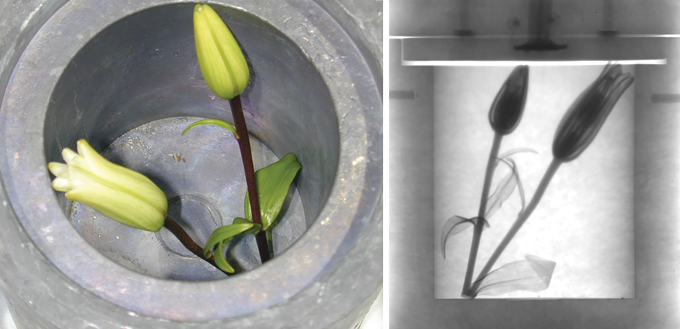
Physicist Jacob LaManna of the Nationwide Institute of Requirements and Era in Gaithersburg, Md., loves to display the comparative features of neutron and X-ray imaging with a CT “nonetheless lifestyles” of Asiatic lilies tucked inside of a hole cask with thick lead partitions. “The neutrons can move throughout the lead, after which you’ll see mainly all of the water [in the] vascular construction of the flora,” LaManna says. An X-ray scan would display not anything however the opaque outer floor of the cask.
The facility to flow via dense fabrics that block X-rays has made neutron imaging a very powerful era for business checking out of cars and planes. The debris can disclose the glide of hydrogen-rich oil inside of engine blocks or reveal flaws in steel castings. For the reason that Nineteen Seventies, U.S. nationwide laboratories have depended on neutron imaging to increase and handle the country’s nuclear guns stockpiles; the neutrons are robust quality-control equipment for mapping out the insides of dense bomb portions and for learning hydrogen-rich fusion explosives inside of warhead elements.
At NIST, LaManna leads the Neutron and X-ray Tomography, or NeXT, facility, which is able to concurrently run X-ray and neutron imaging. The twin perspectives supply distinct but complementary details about issues that include combos of fabrics — like hydrogen gasoline cells, development fabrics and soil samples — that will be tricky to review with just one or the opposite imaging manner.
Over the past couple a long time, as phrase has unfold in regards to the features, a rising collection of paleontologists, archaeologists and anthropologists have added neutron imaging to their analytical toolboxes. Regardless of neutron imaging being round for some time, “we’re in reality the brand new youngsters at the block,” Bevitt says.
Along with revealing a couple of dinosaur bones within the abdominal of a shattered crocodile, along side the femur that to start with stuck Bevitt’s eye, neutron computed tomography has allowed researchers to review the material swaddling cat mummies with out unwrapping them, to find indicators of just lately carried out glues keeping in combination fraudulently assembled artifacts, and discover essentially the most historic vertebrate center ever discovered, in a 380-million-year-old fish.
Rewards and dangers
Paleontologist James Clark puts a couple of fossilized crocodile skulls at the desk in his basement lab at George Washington College in Washington, D.C. The 165-million-year-old fossils are dwarfed by means of a close-by trendy alligator cranium. Whilst the alligator cranium is set so long as my forearm, the fossilized croc skulls are handiest reasonably larger than my thumb tip.
The delicate skulls, which Clark amassed in Mexico 4 a long time in the past, are embedded in hardened blobs of sediment with only a few bones and tooth peeking via. To start with look, the specimens resemble wads of chewed gum, however fabricated from gritty, iron-rich mudstone. “Should you attempt to X-ray that, you mainly finally end up with … those vivid glints from all of the iron,” Clark says. The result’s blurring and streaking that cover the skeletal constructions.
Clark may have employed preparers to scrub away the sediment surrounding the sophisticated bones. But it surely’s a sluggish and dear procedure that may finally end up destructive the specimen, he says.
It wasn’t till 2019 that he in any case were given a excellent have a look at the hidden bones. After a seminar the place he met Bevitt, Clark discovered that neutron scanning might be the solution. The development resulted in an creation to LaManna and the NIST facility 25 kilometers up the street in Maryland.

As a result of iron is basically clear to neutrons, LaManna says, “it’s a lot more uncomplicated to mainly isolate simply the fossil portion of the article.” Photographs from the NIST neutron CT scans printed the intricate main points of the tiny bones. “You’ll be able to then get started enjoying virtual jigsaw puzzles with the bone fragments to take a look at to reconstruct the precise creature.”
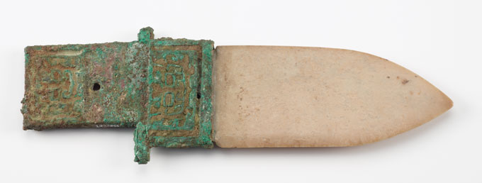
Whilst the fabric round a fossil or object might provide an issue for X-rays, once in a while it’s the article itself that’s the problem. Tissues, fibers, picket and different low-density fabrics will also be tricky to unravel with X-rays, and metals inside of an object can block different options from view. Each demanding situations plague researchers learning antiquities like the three,000-year-old dagger-axes that I noticed on show within the Smithsonian’s Freer Gallery of Artwork in Washington, D.C.
Those ceremonial guns from China’s Shang dynasty are suspended in a vertical glass case, the place I may just get my nostril only a few centimeters from the jade blades and turquoise-encrusted bronze handles. I discovered it was once absolute best to lean up shut in order that I may just respect the intricate blue-green patterns of gems sunk into the steel.
Smithsonian artwork conservator Ariel O’Connor would like to understand how the dagger-axes had been put in combination. X-ray CT doesn’t paintings at the mixture of stone, steel, fibers and different fabrics that can be inside of. Neutron imaging may just lend a hand, but it surely comes with a possibility. Neutron beams make issues radioactive. It’s no longer at all times transparent prematurely how radioactive a pattern will turn out to be, however fabrics steadily exceed the extent of radioactivity that’s protected for people to deal with, and even view in a museum, for days to weeks after publicity to neutron beams.
“Lets in reality do calculations and decide what’s going to be the problematic part and the way lengthy wouldn’t it be radioactive and what kind of,” LaManna says. “[But,] in relation to the jade, the place it’s subject material mainly simply totally dug up from the bottom, it may possibly have all forms of stuff in it that you could no longer essentially be expecting.” That makes residual radioactivity tricky to expect.
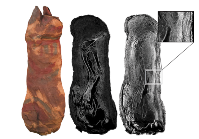
So, O’Connor determined to do a check. She and co-workers made a crude reproduction of an historic dagger-ax. They used jade from Wyoming in lieu of the traditional Chinese language jade, stacks of brass from a repurposed door kickplate to simulate the bronze deal with, and a few silk thread very similar to the kind that holds some Shang dynasty dagger-axes in combination. Then LaManna scanned the dagger with X-rays and neutrons at NIST.
As anticipated, the brass was once fully opaque to the X-rays, hiding options of the reproduction’s development. However the neutron beam printed key main points, together with a view of the jade inserted throughout the brass deal with or even particular person silk threads.
As for residual radioactivity, the reproduction confirmed none of any importance 9 days later. Basically, Bevitt says, residual radiation dies down briefly. One fossil he studied remained radioactive for 3 months, because of the presence of radium, however maximum samples are protected to ship again to labs and museums inside of a couple of weeks or much less.
Nonetheless, with that uncertainty and questions on how chemically an identical the reproduction is to the actual dagger-axes, O’Connor isn’t but in a position to possibility scanning the artifacts.
“As a conservator, I’m entrusted with the preservation and protection of those exceptional 3,000-year-old gadgets to make sure they continue to be for long run generations. If an analytical methodology similar to neutron imaging would possibly resolution our analysis questions however would modify the gadgets and save you them from being out there” because of caused radioactivity, O’Connor says, “we can search for different choices.”
Opening a brand new window to the previous
Regardless of the expanding acclaim for neutron tomography for learning fossils and antiquities, X-ray CT stays the go-to imaging selection for many researchers. Within the Nineteen Nineties, a couple of dozen scholarly papers on the use of neutrons to review the previous had been revealed yearly; just lately, it’s been masses consistent with yr. Publications associated with imaging fossils and artifacts with X-ray CT, despite the fact that, quantity within the 1000’s annually.
More often than not, X-rays suffice, and the benefits are transparent. They provide excessive solution to discover small main points without a lingering radioactivity. X-ray CT machines also are extensively to be had as a result of they’ve been utilized in clinical settings for over 50 years, and so they’re sufficiently small to slot in maximum labs and museum analysis areas.
These days, there are just a few dozen neutron tomography amenities in the world. The particle accelerators and nuclear reactors that produce appropriate neutrons are huge, pricey and closely regulated. Just a handful of the amenities international are to be had to research fossils and antiquities, in line with Burkhard Schillinger, a physicist on the Technical College Munich who runs the neutron imaging beamline there. He ticks off a couple of amenities in america, a part dozen in Europe and one in Australia.
Nonetheless, LaManna says the loss of get entry to doesn’t appear to be the bottleneck in popular adoption of the methodology. Together with the worries over lingering radioactivity, the newness of the era and basic ignorance might stand in the way in which.
“I attempt to recruit as wide a spread of customers as I will” to put up fossils and antiquities for imaging at NIST, LaManna says. “It’s no longer like they’re getting driven out of the way in which” to create space for extra standard neutron research. “It’s simply extra of having the right kind folks to then write proposals, come to us [and] paintings with us to get beam time.”
Within the closing decade, Australia-based Bevitt has unfold the phrase on neutron tomography via lectures and outreach around the globe. Lots of the mavens contacted for this tale hint their preliminary passion in neutron imaging to Bevitt’s affect. Many researchers in his house nation have already embraced the era.
“Principally, in Australia, when a brand new dinosaur is found out,” Bevitt says, “the very first thing that occurs is it involves our lab.”